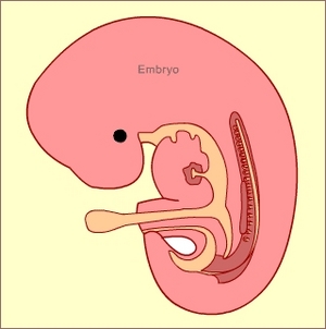
By the time a mother is aware that she is pregnant, she is usually two to three weeks pregnant already. This seems like a short amount of time, but already so much has happened!
Survival of the Fittest
Up to 300 million sperm start out in the race for conception. Out of these, only 1% (3 million) will actually make it to the uterus. These strong few swim upstream and navigate the possibility of two fallopian tubes booby trapped to keep the weak and unsuitable from ever reaching the egg. A mere 500 sperm arrive at the right destination. The strongest few are able to make it through the egg’s protective outer membrane. The first to penetrate the egg wins, and the rest are immediately repelled.
Week One of Fetal Development: Cells Rapidly Divide
Seven days after conception, one sperm and one egg have become several hundred cells. Called a “blastocyst,” this collection of cells has not yet made a home in the uterine lining – it is moving slowly down the fallopian tube and preparing to implant. This is why the pregnancy does not show up yet on a urine test: the hormones that indicate the presence of a fertilized egg won’t activate until implantation takes place.
Dad Is Already the Provider
Both Mom’s genes and Dad’s genes have special chores for helping the development of the fetus. In his book, From First Kicks To First Steps, Dr. Alan Greene writes, “Each cell contains genes from both, but in some cells, more of Dad’s genes are turned on, and in others Mom’s genes are. At this stage, maternal chromosomes are responsible for directing the growth of the budding embryo; paternal chromosomes are responsible for directing the cells that build the surrounding environment – including forging the very placenta that connects mother and child. The father is literally in the middle of this connection” (21).
Week Two of Fetal Development: Implantation
At about 13 days, the blastocyst implants itself in the wall of mother’s uterus and the placenta develops. This organ connects baby to mother. Through it, the embryo (its new name) receives nourishment, and expels wastes for the mom to eliminate with her own.
At the embryo’s end of the placenta, a yolk sac develops. It won’t provide food, since the embryo shares a circulatory system with mom and doesn’t need food storage, but it will develop blood cells until the fetus develops organs that can do this instead (the liver, spleen and bone marrow).
Week Three of Fetal Development: Cells are Assigned Their Jobs
Already various cells are moving to different corners and receiving instructions to become separate organs and body parts such as a skeleton, the digestive system, the circulatory system, or a brain. Before the end of three weeks, a rudimentary nervous system begins to form. This neural center will supervise the rapid fetal development to come.
By day 21, the embryo has a rudimentary spinal column, brain, and heart. The cells that will become eyes appear as disks on top of the “head.” Ear cells are present, but are still waiting to activate.
This tiny, three week old collection of cells has a vascular system that pumps gently around the body; it has, in essence, a heart beat.
From conception to three weeks, the embryo has developed from one cell into millions, and is visible to the naked eye.
Sources:
Green, M.D., Alan. From First Kicks to First Steps: Nurturing Your Baby’s Development from Pregnancy through the First
Year of Life. New York:McGraw-Hill, 2004.
Tsiaras, Alexander and Barry Werth. From Conception to Birth: A Life Unfolds. New York: Doubleday, 2002.
